-
E-mail
sales@caikon.com
-
Phone
13917369745
-
Address
No. 41, Lane 98, Shunda Road, Jiading District, Shanghai (Nanxiang Modern Enterprise Park)
Product Categories
- Metallographic cutting machine QG-4A
- Metallographic cutting machine QG-5A
- Metallographic cutting machine QG-3A
- LED inverted microscope
- Metallographic cutting machine QG-1
- Metallographic cutting machine QG-3
- Metallographic cutting machine QG-5
- Metallographic cutting machine QG-2
- inverted microscope
- hot stage
- Metallographic cutting machine QG-4
- Diamond spray polishing agent
Shanghai Caikang Optical Instrument Co., Ltd

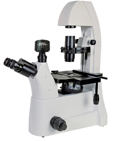
Cai Kang LED Inverted Microscope
NegotiableUpdate on 03/02
- Model
- Nature of the Manufacturer
- Producers
- Product Category
- Place of Origin
Overview
The XDS-700 series inverted microscope combines excellent infinite distance microscopic optical systems with modern high-quality microscopic observation methods, making it easy to achieve observation functions such as bright field, phase contrast, polarized light, and LED fluorescence. It provides an efficient and complete system solution for teaching, research, and detection analysis in fields such as biology and medicine, making microscopic observation easier and more enjoyable.
Product Details
Cai Kang LED Inverted Microscope
1、 The main purpose and characteristics of the instrument | |||||||||||||||||||||||||||||||||||||||||||||||||||||
| |||||||||||||||||||||||||||||||||||||||||||||||||||||
| |||||||||||||||||||||||||||||||||||||||||||||||||||||
| 7、 The principle and application of inverted fluorescence microscope | |||||||||||||||||||||||||||||||||||||||||||||||||||||
|
There are two types of fluorescence microscopes based on their optical paths. 1. Transmission fluorescence microscope: The excitation light source passes through the specimen material through a condenser lens to excite fluorescence. Commonly used dark field concentrators can also be used, and ordinary concentrators can be used to adjust the reflector to redirect and sidelobe the excitation light onto the specimen. This is a relatively old-fashioned fluorescence microscope. Its advantage is strong fluorescence at low magnification, but its disadvantage is that its fluorescence decreases with increasing magnification. Therefore, it is better for observing larger specimen materials. 2. Falling light fluorescence microscope is a new type of fluorescence microscope developed in modern times. Unlike the above, the excitation light falls from the objective lens downwards onto the surface of the specimen, using the same objective lens as the illumination condenser and the objective lens for collecting fluorescence. A dual color beam splitter needs to be added to the optical path, which is 45 degrees apart from the light uranium. Angle, the excitation light is reflected into the objective lens and focused on the sample. The fluorescence generated by the sample, as well as the excitation light reflected from the surface of the objective lens and cover glass, enter the objective lens and return to the dual color beam splitter, separating the excitation light and fluorescence. The residual excitation light is then absorbed by the blocking filter. If different combinations of excitation filters/dual color beam separators/blocking filters are used, they can meet the needs of different fluorescent reaction products. The advantages of this fluorescence microscope are uniform field illumination, clear imaging, and stronger fluorescence with larger magnification. Method of using fluorescence microscope: 1. Turn on the light source, and preheat the ultra-high pressure mercury lamp for a few minutes to reach the maximum bright spot. 2. Transmission fluorescence microscopy requires the installation of the required excitation filter between the lamp source and the condenser, and the corresponding blocking filter behind the objective lens. The falling light fluorescence microscope needs to insert the required excitation filter/dual color beam splitter/blocking filter block into the slot of the optical path. 3. Observe with a low-power microscope, adjust the center of the light source according to the adjustment device of different types of fluorescence microscopes, place the specimen slide, and focus to observe. Attention should be paid during use: Do not observe directly with your eyes before installing the filter to avoid eye damage; When observing specimens with an oil microscope, a special non fluorescent oil microscope must be used; After the high-pressure mercury lamp is turned off, it cannot be immediately turned back on. It takes 5 minutes to restart, otherwise it will be unstable and affect the lifespan of the mercury lamp. (3) Observing cells stained with 0.01% acridine orange fluorescent dye under a fluorescence microscope on a teaching platform using a blue violet filter, two different colors of fluorescence (dark green and orange red) can be observed in the nucleus and cytoplasm. | |||||||||||||||||||||||||||||||||||||||||||||||||||||