-
E-mail
postmaster@ewell.com.cn
-
Phone
18665572216
-
Address
Room 505, Building C1, Information Harbor, Keyun Road, Tianhe Industrial Park, Guangzhou Enterprise QQ: 4009961889
Product Categories
Guangzhou Yuwei Biotechnology Instrument Co., Ltd
Cell imaging system
NegotiableUpdate on 02/25
- Model
- Nature of the Manufacturer
- Producers
- Product Category
- Place of Origin
Overview
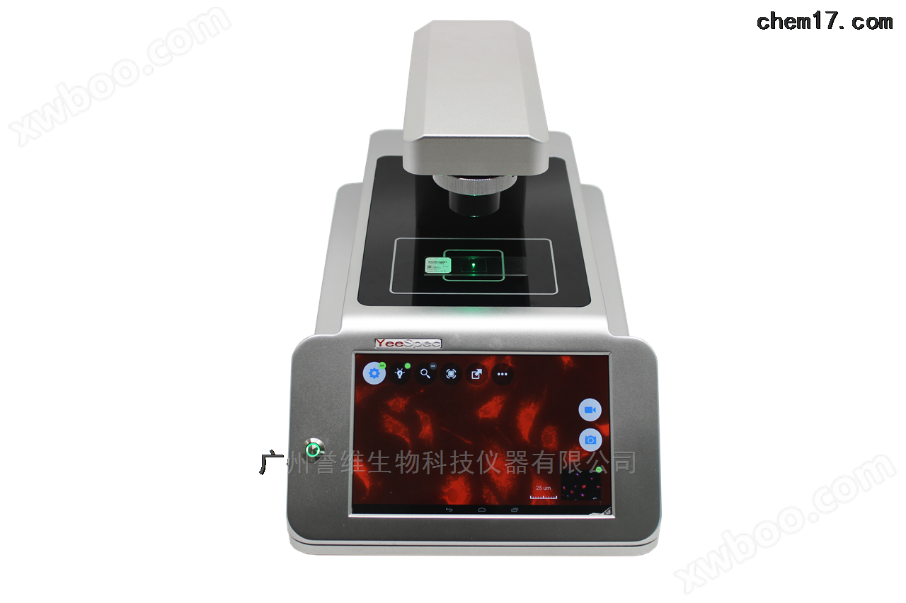
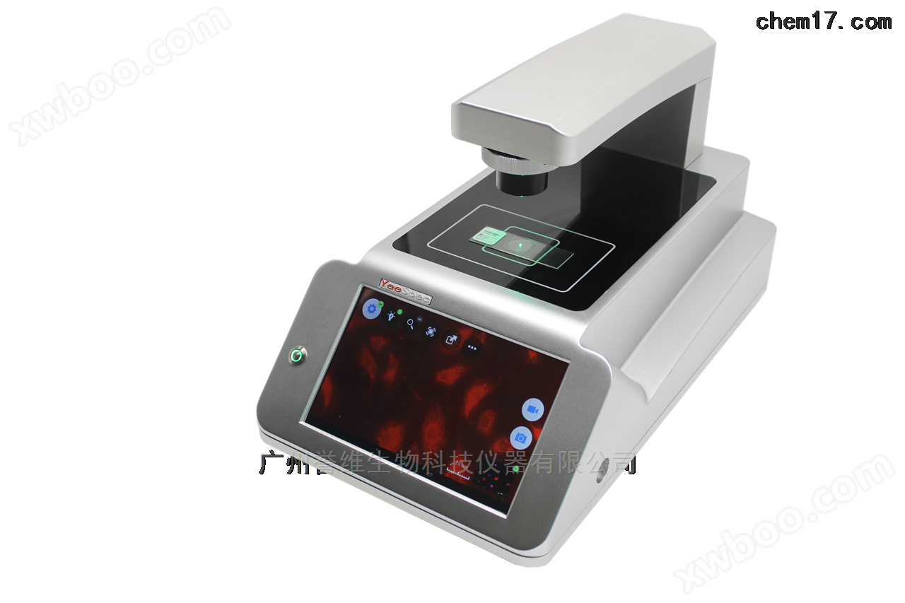
FL Auto 2000s Cell Imaging System is a fast cell imaging observation solution. Whether it's simple cell culture observation, multi-channel imaging observation, or even time series analysis, we can help you quickly complete it
Product Details
Cell imaging systemIt is a method of using optical detection techniques combined with optical detection molecules to image cells, tissues, and even living organisms, in order to obtain biological information. If biological optical imaging is limited to the visible light and near-infrared light range, it can be divided into different types based on the detection methodFluorescence imagingBioluminescence imaging, photoacoustic imaging, optical tomography imaging, etc.
According to the detection method,FL Auto 2000sCell imaging systemFor fluorescence imaging systems,Fluorescence imagingThe technology uses fluorescent reporter groups, including inorganic materials such as upconversion and quantum dots, and organic materials such as green fluorescent protein, red fluorescent protein, or fluorescent dyes for labeling. Using excitation light to achieve a higher molecular energy level of the reporting group, and then emitting longer wavelength visible light to form an in vivo biological light source for detection. The commonly used fluorescent groups currently include various small molecule fluorescent dyes, green fluorescent proteins, and red fluorescent proteins.
working conditions
(1)Environmental temperature:0-40℃
(2)Relative humidity:0-100%RH
(3)Working voltage:100-250V
Purpose:Fast cell imaging, intelligent multi fluorescence channel time series shooting.
Application Direction
(1)Cytotoxicity, screening of cell drugs (lentiviral transfection detection).
(2)Cell apoptosis experiment.
(3)Preliminary screening of protein subcellular localization (multi-channel fluorescence imaging, antigen expression of tumor cells, cellular structural characteristics, effects and mechanisms of anti-tumor drugs, etc.).
(4)Cell culture under low oxygen conditions.
(5)Stem cell monitoring, stem cell proliferation and differentiation.
(6)Optimization of conditions for cell analysis experiments.
(7)Cell Movement and Migration Research (Scratch Analysis)
configuration parameters
| Fluorescence imaging system | |
| model | FL Auto 2000s |
| Number of objective lenses | 2 of them |
| Objective magnification | 10 x phase difference objective lens, |
| 20 x fluorescent objective lens | |
| Instrument memory | 27G |
| Objective switching | electric |
| Objective focusing | Automatic and electric |
| Fluorescence module switching | electric |
| fluorescence channel | 3, highly configurable 4 |
| light source | High brightness solid-state LED light source |
| Fluorescent module | DAPI |
| GFP | |
| RFP | |
| USB port | standard configuration |
| Built-in battery | Large capacity polymer lithium battery (12V, 20000mA) |
| power supply | 110-240V, 50-60Hz, |
| Aviation power socket | |
| software | SpecSee 2.0 |
| time series | have |
| image overlay | have |
| video recording | have |
| Size (L × W × H) | 420×250×252(mm) |
| weight | 16kg |
| Compatible with cultivation containers | Cell culture bottle; Culture dish; Porous plate; Ordinary glass slide |
Application Type
 |
 |
| Osteoblasts determine digestion time, (10x, difference) | Hepatocellular carcinoma cells 20x, DAPI staining |
 |
 |
| Osteosarcoma cell MG63, 20X | Osteosarcoma cell MG63, 20X (blue-green fluorescence overlay) |
 |
|
| 10xRFP fluorescence of ovarian cancer cells |
FL Auto 2000s Fluorescence Cell Imaging, can be placed in a cell culture incubator, * with full touchscreen control and remote operationFluorescence cell imaging