-
E-mail
postmaster@ewell.com.cn
-
Phone
18665572216
-
Address
Room 505, Building C1, Information Harbor, Keyun Road, Tianhe Industrial Park, Guangzhou Enterprise QQ: 4009961889
Product Categories
Guangzhou Yuwei Biotechnology Instrument Co., Ltd
Bright field imaging
NegotiableUpdate on 02/25
- Model
- Nature of the Manufacturer
- Producers
- Product Category
- Place of Origin
Overview
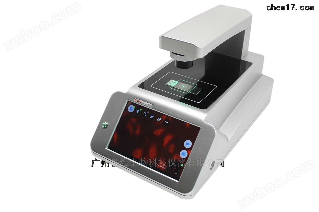
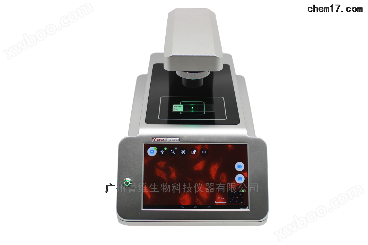
Light Auto bright field imaging, miniature imaging can be placed in cell culture chambers, and full touch screen control supports remote operation. Light Auto magnification supports 5 #215; Flat field objective lens, 10 #215; Phase contrast objective lens; Fully automatic, electric objective focusing; High brightness solid-state LED light source
Product Details
darkBright field imagingPrinciple:The contrast of bright and dark field imaging of crystal thin film samples (i.e. the difference in brightness and darkness in different regions) is formed due to the difference in diffraction intensity caused by the corresponding structure or orientation of different parts of the sample. Therefore, it is called diffraction contrast, and the image formed mainly by the diffraction contrast mechanism is called diffraction contrast.
If only the transmitted beam is allowed to form an image through the objective light barrier, it is called a bright field image; If only a certain diffraction beam is allowed to form an image through the objective light bar, it is called a dark field image.
In terms of diffraction contrast, the differences in structure or orientation in different parts of the sample are actually manifested in the degree to which they satisfy or deviate from the Bragg condition. The region that satisfies the Bragg condition has a higher diffraction beam intensity and a relatively weaker transmission beam intensity, resulting in a dark contrast in the bright field image formed by the transmission beam; On the contrary, the region deviating from the Bragg condition has weaker diffraction beam intensity and relatively higher transmission beam intensity, resulting in bright contrast in the bright field image. The contrast in the dark field image depends on which diffraction beam is selected for imaging. If within a grain, under double beam diffraction conditions, the contrast between the bright field image and the dark field image is exactly opposite.
Dark field imaging is a basic and commonly used technique in transmission electron microscopy, which is relatively easy to operate. Here is a brief introduction to the operation and key points of dark field imaging:
(1) Search for the field of interest under the bright field image.
(2) Insert a selection light bar to surround the selected field of view.
Light AutoBright field imaging
configuration parameters
| model | Light Auto |
| Number of objective lenses | 2 of them |
| Objective magnification | 5 x flat field objective lens, |
| 10 x phase difference objective lens | |
| Instrument memory | 27G |
| Objective switching | electric |
| Objective focusing | Automatic and electric |
| Fluorescence module switching | none |
| fluorescence channel | 1900-01-00 |
| light source | High brightness solid-state LED light source |
| Fluorescent module | none |
| USB port | standard configuration |
| Built-in battery | Large capacity polymer lithium battery (12V, 20000mA) |
| power supply | 110-240V, 50-60Hz, |
| Aviation power socket | |
| software | SpecSee 2.0 |
| time series | have |
| image overlay | none |
| video recording | have |
| Size (L × W × H) | 420×250×252(mm) |
| weight | 15kg |
| Compatible with cultivation containers | Cell culture bottle; Culture dish; Porous plate; Ordinary glass slide |
working conditions
(1) Environmental temperature: 0-40 ℃
(2) Relative humidity: 0-100% RH
(3) Working voltage: 100-250V
Purpose:Fast cell imaging, intelligent multi fluorescence channel time series shooting.
Application Direction
(1) Cytotoxicity, screening of cell drugs (lentiviral transfection detection).
(2) Cell apoptosis experiment.
(3) Preliminary screening of protein subcellular localization (multi-channel fluorescence imaging, antigen expression of tumor cells, cellular structural characteristics, effects and mechanisms of anti-tumor drugs, etc.).
(4) Cell culture under low oxygen conditions.
(5) Stem cell monitoring, stem cell proliferation and differentiation.
(6) Optimization of conditions for cell analysis experiments.
(7) Cell movement and migration research (scratch analysis).

Insect egg sac cells 20x objective lens (bright field observation)