-
E-mail
inquirychina@ohaus.com
- Phone
-
Address
7th Floor, Building 33, No. 680 Guiping Road, Xuhui District, Shanghai
Ohhaus International Trade (Shanghai) Co., Ltd
How to elegantly complete phage amplification and quantitative experiments? Experiment King! |Xiao Ao Classroom
Date: 2022-04-05Read: 0

The start of school season has arrived againExperimental WangWe're going to 'move bricks' again
howExcellentElegant bricklayingstillNo need to seek helpsenior fellow apprentices
Of coursefollowXiao Ao Classroomcolumnla
Today, Xiao Ao will introduce to you
is an articleFull of dry goodsof
Practical Handbook of Phage Display Methods
Amplification and Quantification of Bacteriophages
This is a rare note from a top student
Please keep it in your collection!
Part 1
hybridoma、Single-cell cloningandPhage display technologyThere are three mainstream techniques for antibody discovery, among whichPhage display technologyasNobel Prize level technologyIt plays a very important role in the process of biological innovation and pharmaceutical research and development,*The acceleration of the development process of antibody drugs.Phage display technologyNot only does it play a core role in the discovery and optimization of new antibodies and peptides, but it can also be combined with traditional animal immune technology and effectively docked with nanobodies and fully human transgenic mouse discovery platforms,Can be widely used in the fields of antibodies, antibody coupled drugs, and CAR-T/TCR-T.This article will explore the details of phage proliferation and amplification experiments in phage display technology, share specific experimental process experiences, and guide the experimental process based on its principles. With a focus on excellence in the experimental process, the success rate of phage display technology in antibody discovery will be increased.
Part 2
——Phage library
Basic principles of amplification and display|
The widely used phage display system for antibody fragments is called3+3 system(3 is the P3 capsid protein of bacteriophage);3+3The P3 capsid protein has two sources:
- Phagemids
- M13KO7 Helper Phage
Bacterial granulesThe P3 protein on phage fused with the gene of the antibody fragment is the key to library diversity;Auxiliary bacteriophageThe P3 protein on Helper Phage is the natural P3 protein of bacteriophages.
*Phagophages are relatively small plasmid vectors that have high transformation efficiency in the competent state of Escherichia coli and can be amplified in E. coli.
After being infected with helper phages, Escherichia coli containing phages will utilize the enzyme system, raw materials, and energy of E. coli for amplification. Eventually, the phages containing phages will be released from E. coli, and the P3 capsid protein of the progeny phages will display antibody fragments.
Part 3
——During phage amplification process
Difficulties encountered and corresponding solutions|
The process of constructing phage libraries is actually the transmission of antibody fragment gene diversity:
The process of transferring the diversity of antibody genes from blood samples to phagemids is the content of constructing phagemid libraries;
The transfer of diversity from phagemids to E. coli containing phagemids is the content of phagemid plasmid electroporation of E. coli;
The transfer of diversity from Escherichia coli containing bacteriophages to bacteriophages is the focus of bacteriophage amplification and the experimental experience in this article.
The ultimate goal of phage amplification is to convert a single copy of the antibody fragment gene in the bacteriophage into 10-100 copies through the phage amplification process,And these copied genes can display antibody protein fragments on the P3 protein on the surface of bacteriophages.
In order to maintain and transfer the diversity of the library during the amplification process of bacteriophages, it is necessary to determine the number and volume of Escherichia coli containing bacteriophages during the amplification process based on the initial size of the library, the corresponding number of auxiliary bacteriophages added for effective infection, and the number of bacteriophages containing bacteriophages added for selection after amplification, which need to be calculated and evaluated one by one,Bacteriophage libraries of different library sizes require different amplification volumes during the amplification process.A fixed experimental plan is likely to lead to the loss of library diversity.
To clarify these issues, one must first have the following basic prerequisite knowledge:
( 👇 The following content is all highlighted!!! )
1. The reading of TG1 Escherichia coli at OD600: 1OD represents a bacterial concentration of 1e9 cells/ml.
2. Phages/helper phages have a relatively good infection efficiency when TG1 is in logarithmic growth mode, and the OD600 value of TG1 in logarithmic growth mode is between 0.4-0.8.
3. The growth rate of TG1 in logarithmic growth is 20 minutes per generation, and it is necessary to closely monitor the growth of TG1 and timely collect bacteria for experiments.4. The MOI ratio of helper phages to TG1 at 20:1 can ensure that all TG1 are infected.
5. Bacteriophages containing bacteriophages can infect normal TG1 Escherichia coli, leaving the bacteriophages in TG1 and amplifying them with the amplification of TG1. In the absence of auxiliary bacteriophages, there is no bacteriophage amplification.
6.Phagophage plasmids contain * resistance, while helper phages contain * resistance.
7. Bacteriophages containing bacteriophages can be titrated by infecting normal TG1 and gradient dilution onto resistant agar plates.8.In the 3+3 mode, bacteriophages containing phagemids exhibit relatively low efficiency in displaying P3 fusion antibody fragment proteins, only 5% -10%. During selection, the titer of the added bacteriophages should be more than 100 times the diversity of the library.
Another crucial issue isBacteriophages, as a type of virus, can easily spread into the air in the form of aerosols, contaminating the laboratory environment and infecting factor F positive Escherichia coli.(Heartfelt for the biological dog, risking his life to move bricks!)
InExperiments related to phage displayNeed to be conducted in a relatively closed laboratoryTo avoid contamination of Escherichia coli by bacteriophages,Especially when cultivating normal TG1 Escherichia coli. The inactivation method of bacteriophages is usually UV irradiation for more than half an hour.(A qualified biological dog, a virus killer without emotions!)
Part 4
——Bacteriophage expansionIncrease and quantify
Sharing of specific experimental process|
The following is an experimental protocol for amplifying a phage library with a library size of 1E9.
*If the library is too large or too small, the amplification volume can be increased or decreased proportionally.
Day 1(A Bald Day Moving Bricks)
1. Take 1ml of TG1 strain containing bacteriophages (with a bacterial count of 1E10) and add it to a 250ml shake flask containing 100ml of 2xYT-GA medium,useOhhaus shakerShake the bacterial shaker at 37 degrees and 220rpm until the OD600 is approximately 0.5.2. Take 20ml of bacterial solution with an OD600 of approximately 0.5 (number of bacteria is 1E10), add auxiliary bacteriophage M13 K07 with a pfu of 2E11 according to a MOI of 20:1, and shake at 37 degrees Celsius and 220rpm for half an hour for infection.3. Take the infected bacterial solution and transfer it to a 50ml centrifuge tube for useOhhaus centrifuge 5816RCentrifuge 3200g for 10 minutesRemove the culture medium to eliminate the inhibition of P3 protein fusion antibody fragment protein expression by glucose in the culture medium.4. Resuspend the bacterial colony in 2xYT-AK medium, add 400ml of medium, and shake overnight at 30 ° C and 220rpm in a 1L shake flask.
Day 2(The second day after moving bricks and becoming bald)
5.Divide 400ml of bacterial solution evenly into large centrifuge bottles,useOhhaus centrifuge 5816R Centrifuge 10000g for 10 minutesTake the supernatant, centrifuge 10000g again for 10 minutes,Remove residual bacterial fragments.6. Add 90ml of PEG solution to 400ml of supernatant, incubate at 4 ℃ for 1 hour with gentle shaking, and thenUsing a device with low-temperature control functionOhhaus centrifuge 5816R Centrifuge at 4 ℃ 10000g for 1 hour.7. Remove the supernatant, resuspend the bacteriophage with 10ml PBS, and add 2.5ml PEG solution for further precipitation8. Incubate on ice for 20 minutes, thenUsing a device with low-temperature control functionOhhaus centrifuge 5816RCentrifuge at 4 ℃ 10000g for half an hour.9. Remove the supernatant, use absorbent paper to absorb the residual PEG solution, add 1-2ml of PBS to resuspend the bacteriophage, and perform titer determination.
Titer determination:
1. In a separate laboratory, normal TG1 Escherichia coli was taken and added to 2xYT culture medium,useOhhaus shakerShake the bacteria to an OD600 value of 0.5If not used in a timely manner, it can be stored at 4 ℃ for 2 hours as a backup.
2.Dilute the amplified phage solution in step 9 with PBS at a ratio of 1E5-1E7 times.
3. Add 10ul of phage dilution solution to 490ul of TG1 Escherichia coli with an OD600 of 0.5,useOhhaus shaker 37 ℃ 220rpm oscillation for half an hourCarry out infection.
4. Take 50ul of infection solution and evenly apply it to an agar plate containing 2xYT-GA. After drying, place it in a 37 ℃ incubator overnight. And apply non infected TG1 E. coli onto agar plates as a negative control.
Day 3On the third day of moving bricks and becoming bald
5. Calculate the number of clones in a 2xYT-GA agar plate and determine the titer of the amplified bacteriophage based on the dilution factor
Phage titer (pfu/ml)=(number of clones * 10 * 50/10ul) * dilution factor
6.Take bacteriophages from 1E11-1E12 for selection.
Reagent formula:
2xYT medium:Yeast Extract 10.0 g/L+Tryptone 16.0 g/L+NaCl 5.0 g/L
2xYT-GA medium:2xYT medium+100 μ g/ml *+1% (W/V) glucose
2xYT-AK medium:2xYT medium+100 μ g/ml *+50 μ g/ml*
PEG溶液:20% (w/v) PEG 6000, 2.5 M NaCl
Part 5
Instrument *:
Every day as a research worker, I am not alone because I have Xiao Ao by my side
Bacteriophages, as a type of virus, can easily spread into the air in the form of aerosols, polluting the laboratory environment. A closed laboratory is required, and all laboratory instruments must be used separately.
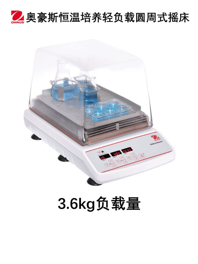
ohausConstant temperature light load cultivation shakerThe speed range is 100-12-rpm, with a compact size and powerful functions, providing excellent shaking effect for your samples:
In addition, during the phage display experiment, there will be centrifugation steps with different volumes, speeds, and requiring 4 ℃ conditions. Ifwithmany
Centrifugal machineMeet the needs of the experimentIt will result in the vacancy of experimental equipment assets and cause * waste!!!
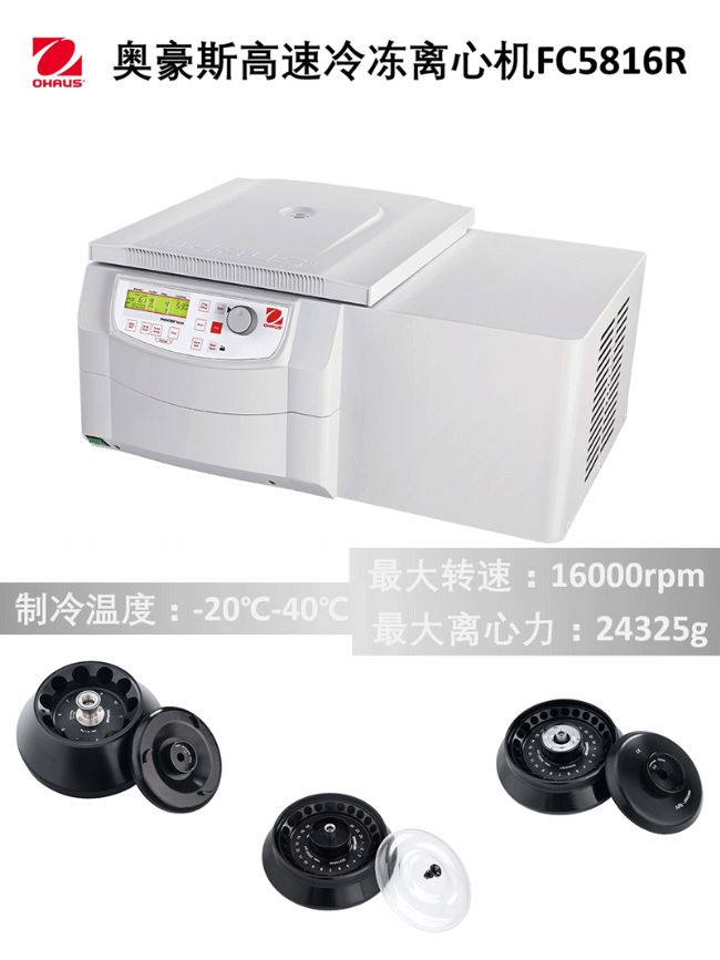
ohaus5816R multifunctional centrifugeThe optional rotor provides convenience for bacteriophage related experiments, and one centrifuge meets all experimental needs!
Next, Xiao Ao will take you on a rideThe car that helps you save research fundingTeach you how to properly pluck the wool of Ohouse
Germany*
Powerful functionality and excellent centrifugal effect
O'Hare Frontier ™ Centrifuge family
launchBuy one get one free "*Activity
*The model is as follows
Micro high-speed centrifugeFC5515R(Frozen model)
Micro high-speed centrifugeFC5515(Non frozen model)
New compact micro centrifugeFC5513
Get in the car now, there's still plenty of time!
Please stamp the poster for details and scan the QR code for registration 👇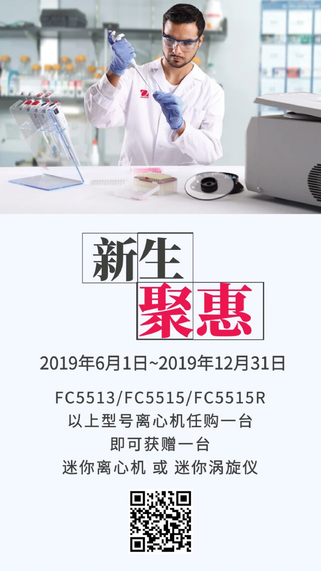
Want to learn more aboutOhhaus Laboratory Productsinformation? Please enterRead the full textLeave a message, our professional engineers will come quickly to serve you!
▼